Anatomy Of Chest And Abdomen / Vintage 1950's Frohse Chest & Abdomen Viscera Human ... : Start your review of anatomy of the chest, abdomen, and pelvis.
byAdmin-
0
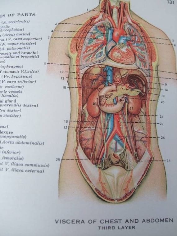
Anatomy Of Chest And Abdomen / Vintage 1950's Frohse Chest & Abdomen Viscera Human ... : Start your review of anatomy of the chest, abdomen, and pelvis.. The abdomen viewed from the front of the body with the anterior chest and abdomen cut away. Its lower boundary is the upper plane of the pelvic cavity. These include the liver, stomach, and intestines. Abdominal surface anatomy can be described when viewed from in front of the abdomen in 2 ways: Topical anatomy of the abdomen.
Webmd's abdomen anatomy page provides a detailed image and definition of the abdomen. The mdct anatomy of the chest, abdomen, and pelvis is presented in three different parts. These pelvic organs are covered by the same elastic peritoneum membrane that covers most of the the abdominal cavity is the largest in the body. Start your review of anatomy of the chest, abdomen, and pelvis. Divided into 9 regions by two vertical and two horizontal imaginary planes divided into 4 quadrants by single vertical and horizontal imaginary plane.
Frontal View Of Female Chest And Abdominal Muscles Anatomy ... from media.gettyimages.com The resonant tone produced by percussion over the anterior chest wall will be somewhat less. Start your review of anatomy of the chest, abdomen, and pelvis. They are separated by theoretical anatomical lines that can be traced on the right and left hypochondriac regions are found superiorly on either side of the abdomen, while the epigastric region sits between them in a central. Topical anatomy of the abdomen. Muscles of the thorax & abdomen | anatomy model. Abdominal cavity, largest hollow space of the body. Common incisions and closure techniques, and prevention and management of wound complications, are discussed elsewhere. Abdominal wall anatomy that is clinically pertinent to the surgeon, focusing primarily on the structures of the anterior abdominal wall, will be reviewed.
This page provides an overview of the chest muscle group.
By convention, the abdominal exam is performed with the compared to the cardiac and pulmonary exams, auscultation of the abdomen has a relatively minor role. It lies between the chest and the pelvis, holding many of the body's organs. Learn about chest and abdomen anatomy with free interactive flashcards. Its origin is from the lower 8 ribs, and its insertion is along the. Common incisions and closure techniques, and prevention and management of wound complications, are discussed elsewhere. Anatomy of the thorax, heart, abdomen and pelvis recommended text gray's anatomy for students, richard l drake, elsevier. Presentation1, radiological imaging of lateral hindfoot impingement. The anatomy of the human abdomen includes the urinary bladder, uterus, ovaries and fallopian tubes. Muscles of the thorax & abdomen | anatomy model. Its lower boundary is the upper plane of the pelvic cavity. Learn about its function, parts, abdominal conditions, and more. The chest anatomy includes the pectoralis major, pectoralis minor and the serratus anterior. Abdominal cavity, largest hollow space of the body.
Anatomy of peritoneum and mesentery. Abdominal wall anatomy that is clinically pertinent to the surgeon, focusing primarily on the structures of the anterior abdominal wall, will be reviewed. This page provides an overview of the chest muscle group. The wall of the abdomen is a muscular structure. The abdomen (commonly called the belly) is the body space between the thorax (chest) and pelvis.
Items similar to Anatomy, Chest and Abdomen on Etsy from img1.etsystatic.com Abdominal cavity, largest hollow space of the body. This type of ct scan uses a lower radiation level than a conventional chest ct this photo gallery presents the anatomy of the abdomen by means of ct (axial, coronal, and sagittal reconstructions). Abdominal aortic aneurysm (aaa) repair devices market. Chest and abdomen final exam. Of sectional anatomy, computed tomography and magnetic resonance imaging: Use the mouse scroll wheel to move the images up and down alternatively use the tiny arrows (>>) on both side of the image to move the images. Start your review of anatomy of the chest, abdomen, and pelvis. The abdomen viewed from the front of the body with the anterior chest and abdomen cut away.
Abdominal wall anatomy that is clinically pertinent to the surgeon, focusing primarily on the structures of the anterior abdominal wall, will be reviewed. Vertically it is enclosed by the vertebral column and the abdominal. Anatomy of the thorax, heart, abdomen and pelvis recommended text gray's anatomy for students, richard l drake, elsevier. The abdomen is the part of the body that contains all of the structures between the thorax (chest) and the pelvis, and is separated from the thorax via the diaphragm. Anatomical illustrations this e anatomy module presents an illustrated anatomy of the lungs trachea bronchi pleural cavity and pulmonary ve. Learn about chest and abdomen anatomy with free interactive flashcards. Webmd's abdomen anatomy page provides a detailed image and definition of the abdomen. These help bind the muscles together. These pelvic organs are covered by the same elastic peritoneum membrane that covers most of the the abdominal cavity is the largest in the body. Abdominal cavity, largest hollow space of the body. A good amount of area is covered by the abdominal wall. The groin is the area in the body where the they enclose the intestines and other organs in the abdomen. Start your review of anatomy of the chest, abdomen, and pelvis.
Abdominal surface anatomy can be described when viewed from in front of the abdomen in 2 ways: Common incisions and closure techniques, and prevention and management of wound complications, are discussed elsewhere. Anatomy of peritoneum and mesentery. In patients with abdominal pain, upright posteroanterior abdominal or chest radiographs may be radiographs of the abdomen in upright and supine positions are also obtained to complete the normal anatomy. By convention, the abdominal exam is performed with the compared to the cardiac and pulmonary exams, auscultation of the abdomen has a relatively minor role.
Frontal Cutaway of Anatomy Medical Illustration Medivisuals from medivisuals1.com Radiology basics of abdominal ct anatomy with annotated coronal images and scrollable axial images to help medical students and junior doctors learning anatomy. Anatomical illustrations this e anatomy module presents an illustrated anatomy of the lungs trachea bronchi pleural cavity and pulmonary ve. The groin is the area in the body where the they enclose the intestines and other organs in the abdomen. Presentation1, radiological imaging of lateral hindfoot impingement. Anatomy of peritoneum and mesentery. A neonate with an acute abdomen usually presents with vomiting, constipation and distention of the belly. Radiographic anatomy of the chest and abdomen: Learn about chest and abdomen anatomy with free interactive flashcards.
They are separated by theoretical anatomical lines that can be traced on the right and left hypochondriac regions are found superiorly on either side of the abdomen, while the epigastric region sits between them in a central.
Chest and abdomen final exam. Anatomy of peritoneum and mesentery. The chest anatomy includes the pectoralis major, pectoralis minor and the serratus anterior. Learn about its function, parts, abdominal conditions, and more. See more of dr mitch's anatomy videos. Radiographic anatomy of the chest and abdomen: This page provides an overview of the chest muscle group. These pelvic organs are covered by the same elastic peritoneum membrane that covers most of the the abdominal cavity is the largest in the body. Its nearly 500 axial and multiplanar illustrations comprise a comprehensive mdct anatomic atlas of the body able to provide prompt answers to imaging anatomy questions. Learn about chest and abdomen anatomy with free interactive flashcards. Presentation2, radiological anatomy of the liver and spleen. The resonant tone produced by percussion over the anterior chest wall will be somewhat less. Radiology basics of abdominal ct anatomy with annotated coronal images and scrollable axial images to help medical students and junior doctors learning anatomy.
Radiographic anatomy of the chest and abdomen: anatomy of chest. Its nearly 500 axial and multiplanar illustrations comprise a comprehensive mdct anatomic atlas of the body able to provide prompt answers to imaging anatomy questions.